
Development and impact of histoguide application towards drawing and labelling in microscopic practical
DOI:
https://doi.org/10.31129/LUMAT.12.4.2407Keywords:
HistoGuide, virtual microscopy, validity, usability, drawingAbstract
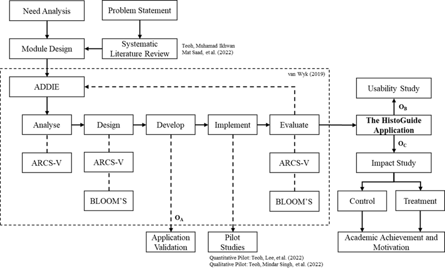
The HistoGuide is an Android application used for virtual microscopy and slides to solve the problems of incorrect drawing and labelling in microscopic practicals. It is developed based on modified Analyze, Design, Develop, Implement, and Evaluate (ADDIE), the van Wyk model, as a self-regulated mobile learning, complementary to optical microscopy. However, as a newly developed application, many still do not understand the usability and impact of virtual microscopy. Hence, the HistoGuide was validated using Cohen’s kappa agreement coefficient, strengthened with the Content Validity Index (CVI). Data were analysed descriptively using mean, standard deviation and percentages for the usability study and inferentially using independent and paired sample t-tests for the impact study. Findings revealed that Cohen’s kappa for content, pedagogy, and technology constructs are 1.00, 1.00, and 0.90, respectively, with an overall of 0.96. The HistoGuide application also achieved high I-CVI and excellent content validity of the overall validation with S-CVI/UA of 0.80 and S-CVI/Ave of 0.96. As for the usability study, the HistoGuide application recorded a high usability level for the overall usability and its four usability constructs: usefulness, ease of use, ease of learning and satisfaction. In the assessment achievement study, there were significant differences between pre- and post-test scores for the treatment group and post-test scores between the treatment and control groups. Thus, the treatment group performed very well compared to the control group in terms of assessment achievement. In the motivation study, the treatment group performed better than the control in motivation and its five motivation constructs: attention, relevance, confidence, satisfaction, and volition. Overall, students from the treatment group outperformed in assessment achievement and motivation compared to the control after using the HistoGuide application. HistoGuide application could enhance the drawing and labelling based on the usability and impact study. This study implies that virtual microscopy could promote innovative learning of microscopic practicals.
References
Bacha, D., Ferjaoui, W., Charfi, L., Rejaibi, S., Slama, S. Ben, Njim, L., & Lahmar, A. (2020). The interest of virtual microscopy as a means of simulation learning in pathological anatomy and cytology. Onkologia i Radioterapia, 14(5), 23–29.
Cheng, X., Lee, K. K. ho, Chang, E. Y., & Yang, X. (2017). The “flipped classroom” approach: Stimulating positive learning attitudes and improving mastery of histology among medical students. Anatomical Sciences Education, 10(4), 317–327. https://doi.org/10.1002/ase.1664 DOI: https://doi.org/10.1002/ase.1664
Cheung, K. K. C., & Winterbottom, M. (2021). Exploring students’ visualisation competence with photomicrographs of villi. International Journal of Science Education, 43(14), 2290–2315. https://doi.org/10.1080/09500693.2021.1959958 DOI: https://doi.org/10.1080/09500693.2021.1959958
Chua, Y. P. (2020). Mastering research statistics (2nd ed.). McGraw-Hill Education (Malaysia).
Cohen, J. (1960). A coefficient of agreement for nominal scales. Educational and Psychological Measurement, 20(1), 37–46. DOI: https://doi.org/10.1177/001316446002000104
Cromley, J. G., Perez, T., Kaplan, A., Dai, T., Mara, K., & Balsai, M. J. (2020). Combined cognitive-motivational modules delivered via an LMS increase undergraduate biology grades. Technology, Mind, and Behavior, 1(2), 1–19. https://doi.org/10.1037/tmb0000020.supp DOI: https://doi.org/10.1037/tmb0000020
Dickerson, J., & Kubasko, D. (2007). Digital microscopes: Enhancing collaboration and engagement in science classrooms with information technologies. Contemporary Issues in Technology and Teacher Education, 7(4), 279–292.
Donnelly, A. D., Mukherjee, M. S., Lyden, E. R., & Radio, S. J. (2012). Virtual microscopy in cytotechnology education: Application of knowledge from virtual to glass. CytoJournal, 9(1). https://doi.org/10.4103/1742-6413.95827 DOI: https://doi.org/10.4103/1742-6413.95827
Fatimah Mohamed, Tan, S. W., & Noor, N. N. M. (2011). Observing and sketching skills in plant anatomy practical class. Jurnal Sains Dan Matematik, 3(2), 66–73.
García, M., Victory, N., Navarro-Sempere, A., & Segovia, Y. (2019). Students’ views on difficulties in learning histology. Anatomical Sciences Education, 12(5), 541–549. https://doi.org/10.1002/ase.1838 DOI: https://doi.org/10.1002/ase.1838
Giannakas, F., Kambourakis, G., Papasalouros, A., & Gritzalis, S. (2018). A critical review of 13 years of mobile game-based learning. Educational Technology Research and Development, 66(2), 341–384. https://doi.org/10.1007/s11423-017-9552-z DOI: https://doi.org/10.1007/s11423-017-9552-z
Hamidah Yusof, Jamal Yunus, & Khalip Musa. (2015). Kaedah penyelidikan : Pengurusan pendidikan. Penerbit Universiti Pendidikan Sultan Idris.
Hande, A. H., Lohe, V. K., Chaudhary, M. S., Gawande, M. N., Patil, S. K., & Zade, P. R. (2017). Impact of virtual microscopy with conventional microscopy on student learning in dental histology. Dental Research Journal, 14(2), 111–116. https://doi.org/10.4103/1735-3327.205788 DOI: https://doi.org/10.4103/1735-3327.205788
Hariyanto, D., Triyono, M. B., & Köhler, T. (2020). Usability evaluation of personalised adaptive e-learning system using USE questionnaire. Knowledge Management and E-Learning, 12(1), 85–105. https://doi.org/10.34105/j.kmel.2020.12.005 DOI: https://doi.org/10.34105/j.kmel.2020.12.005
Helle, L., Nivala, M., Kronqvist, P., Gegenfurtner, A., Björk, P., & Säljö, R. (2011). Traditional microscopy instruction versus process-oriented virtual microscopy instruction: A naturalistic experiment with control group. Diagnostic Pathology, 6(8), 1–9. https://doi.org/10.1186/1746-1596-6-S1-S8 DOI: https://doi.org/10.1186/1746-1596-6-S1-S8
Keller, J. M. (2010). Motivational design for learning and performance: The ARCS model approach. Springer. DOI: https://doi.org/10.1007/978-1-4419-1250-3
Keller, J. M., Ucar, H., & Kumtepe, A. T. (2020). Development and validation of a scale to measure volition for learning. Open Praxis, 12(2), 161–174. https://doi.org/10.5944/openpraxis.12.2.1082 DOI: https://doi.org/10.5944/openpraxis.12.2.1082
Krippendorf, B. B., & Lough, J. (2005). Complete and rapid switch from light microscopy to virtual microscopy for teaching medical histology. Anatomical Record - Part B New Anatomist, 285(1), 19–25. https://doi.org/10.1002/ar.b.20066 DOI: https://doi.org/10.1002/ar.b.20066
Kumar, R. K., Velan, G. M., Korell, S. O., Kandara, M., Dee, F. R., & Wakefield, D. (2004). Virtual microscopy for learning and assessment in pathology. Journal of Pathology, 204(5), 613–618. https://doi.org/10.1002/path.1658 DOI: https://doi.org/10.1002/path.1658
Lavasani, M. G., Mirhosseini, F. S., Hejazi, E., & Davoodi, M. (2011). The effect of self-regulation learning strategies training on the academic motivation and self-efficacy. Procedia - Social and Behavioral Sciences, 29, 627–632. https://doi.org/10.1016/j.sbspro.2011.11.285 DOI: https://doi.org/10.1016/j.sbspro.2011.11.285
Lockee, B. B. (2021). Online education in the post-COVID era. Nature Electronics, 4, 5–6. https://doi.org/10.1038/s41928-020-00534-0 DOI: https://doi.org/10.1038/s41928-020-00534-0
Loorbach, N., Peters, O., Karreman, J., & Steehouder, M. (2015). Validation of the Instructional Materials Motivation Survey (IMMS) in a self-directed instructional setting aimed at working with technology. British Journal of Educational Technology, 46(1), 204–218. https://doi.org/10.1111/bjet.12138 DOI: https://doi.org/10.1111/bjet.12138
Lund, A. M. (2001). Measuring usability with the USE questionnaire. Usability Interface, 8(2), 3–6.
Manganello, F., Falsetti, C., & Leo, T. (2019). Self-regulated learning for web-enhanced control engineering education. Educational Technology and Society, 22(1), 44–58.
Mojica, E. R. E., & Upmacis, R. K. (2022). Challenges encountered and students’ reactions to practices utilised in a general chemistry laboratory course during the COVID-19 pandemic. Journal of Chemical Education, 99(2), 1053–1059. https://doi.org/10.1021/acs.jchemed.1c00838 DOI: https://doi.org/10.1021/acs.jchemed.1c00838
Nauhria, S., & Hangfu, L. (2019). Virtual microscopy enhances the reliability and validity in histopathology curriculum: Practical guidelines. MedEdPublish, 8, 28. https://doi.org/10.15694/mep.2019.000028.2 DOI: https://doi.org/10.15694/mep.2019.000028.1
Neel, J. A., Grindem, C. B., & Bristol, D. G. (2007). Introduction and evaluation of virtual microscopy in teaching veterinary cytopathology. Journal of Veterinary Medical Education, 34(4), 437–444. https://doi.org/10.3138/jvme.34.4.437 DOI: https://doi.org/10.3138/jvme.34.4.437
Ordi, O., Bombí, J. A., Martínez, A., Ramírez, J., Alòs, L., Saco, A., Ribalta, T., Fernández, P. L., Campo, E., & Ordi, J. (2015). Virtual microscopy in the undergraduate teaching of pathology. Journal of Pathology Informatics, 6(1), 1–6. https://doi.org/10.4103/2153-3539.150246 DOI: https://doi.org/10.4103/2153-3539.150246
Polit, D. F., Beck, C. T., & Owen, S. V. (2007). Focus on research methods: Is the CVI an acceptable indicator of content validity? Appraisal and recommendations. Research in Nursing & Health, 30, 459–467. https://doi.org/10.1002/nur.20199 DOI: https://doi.org/10.1002/nur.20199
Raja, S. (2010). Virtual microscopy as a teaching tool adjuvant to traditional microscopy. Medical Education, 44(11), 1126. https://doi.org/10.1111/j.1365-2923.2010.03841.x DOI: https://doi.org/10.1111/j.1365-2923.2010.03841.x
Rodrigues, I. B., Adachi, J. D., Beattie, K. A., & MacDermid, J. C. (2017). Development and validation of a new tool to measure the facilitators, barriers and preferences to exercise in people with osteoporosis. BMC Musculoskeletal Disorders, 18(1), 1–9. https://doi.org/10.1186/s12891-017-1914-5 DOI: https://doi.org/10.1186/s12891-017-1914-5
Saco, A., Bombi, J. A., Garcia, A., Ramírez, J., & Ordi, J. (2016). Current status of whole-slide imaging in education. Pathobiology, 83(2–3), 79–88. https://doi.org/10.1159/000442391 DOI: https://doi.org/10.1159/000442391
Shi, J., Mo, X., & Sun, Z. (2012). Content validity index in scale development. Journal of Central South University (Medical Sciences), 37(2), 152–155. https://doi.org/10.3969/J.ISSN.1672-7347.2012.02.007
Simok, A. A., Hadie@Haji, S. N. H., Abdul Manan@Sulong, H., Yusoff, M. S. B., Mohd Noor, N. F., Asari, M. A., & Kasim, F. (2019). The impact of virtual microscopy on medical students’ intrinsic motivation. Education in Medicine Journal, 14(4), 47–59. https://doi.org/10.21315/eimj2019.11.4.5 DOI: https://doi.org/10.21315/eimj2019.11.4.5
Tauber, Z., Cizkova, K., Lichnovska, R., Lacey, H., Erdosova, B., Zizka, R., & Kamarad, V. (2019). Evaluation of the effectiveness of the presentation of virtual histology slides by students during classes. Are there any differences in approach between dentistry and general medicine students? European Journal of Dental Education, 23(2), 119–126. https://doi.org/10.1111/eje.12410 DOI: https://doi.org/10.1111/eje.12410
Teoh, C. Z., Lee, T. T., Muhamad Ikhwan Mat Saad, Sagat, M. P., Nur Aqilah Jamaludin Khir, & Rajagopal, T. (2022). HistoGuide mobile application (virtual microscopy and slides): A quantitative usability pilot study. Journal of ICT in Education, 9(2), 162–174. https://doi.org/10.37134/jictie.vol9.2.12.2022 DOI: https://doi.org/10.37134/jictie.vol9.2.12.2022
Teoh, C. Z., Mindar Singh, N. K., Siti Nor Badariah, & Marzuki Mohd Uzi Dollah. (2022). Using HistoGuide mobile application (virtual microscopy): A qualitative pilot study on usability and sixth form students’ learning experience. EDUCATUM Journal of Science, Mathematics and Technology, 9(2), 136–154. https://doi.org/10.37134/ejsmt.vol9.2.15.2022 DOI: https://doi.org/10.37134/ejsmt.vol9.2.15.2022
Teoh, C. Z., Muhamad Ikhwan Mat Saad, & Che Nidzam Che Ahmad. (2022). Integrating technology-mediated learning in biology education (histology): A systematic literature review. Journal of ICT in Education, 9(1), 86–99. https://doi.org/10.37134/jictie.vol9.1.8.2022 DOI: https://doi.org/10.37134/jictie.vol9.1.8.2022
Triola, M. M., & Holloway, W. J. (2011). Enhanced virtual microscopy for collaborative education. BMC Medical Education, 11(1), 9–12. https://doi.org/10.1186/1472-6920-11-4 DOI: https://doi.org/10.1186/1472-6920-11-4
van Wyk, N. (2019). A proposed framework for developing online courses: The case for a VR course. University of Cape Town.
van Wyk, N., Johnston, K., Moeller, K., & Haas, F. (2020). Developing an IT course for emerging technologies using a framework – An example of an IoT Course V1.0. Proceedings of the 2020 InSITE Conference, 015–045. https://doi.org/10.28945/4521 DOI: https://doi.org/10.28945/4521
Xu, C. J. (2013). Is virtual microscopy really better for histology teaching? Anatomical Sciences Education, 6(2), 138. https://doi.org/10.1002/ase.1337 DOI: https://doi.org/10.1002/ase.1337
Zamanzadeh, V., Ghahramanian, A., Rassouli, M., Abbaszadeh, A., Alavi-Majd, H., & Nikanfar, A.-R. (2015). Design and implementation content validity study: Development of an instrument for measuring patient-centered communication. Journal of Caring Sciences, 4(2), 165–178. https://doi.org/10.15171/jcs.2015.017 DOI: https://doi.org/10.15171/jcs.2015.017
Downloads
Published
How to Cite
Issue
Section
Categories
License
Copyright (c) 2025 Muhamad Ikhwan Mat Saad, Chern Zhong Teoh , Mohd Mokhzani Ibrahim, Mohamad Termizi Borhan, Eng Tek Ong, Mohd Afifi Bahurudin Setambah

This work is licensed under a Creative Commons Attribution 4.0 International License.


